


o r t h o p a e d i c s . c o . u k

| +home | |
| +news | |
| +research | |
| +patient infomation | |
| +the clinic | |
| +the surgeon | |
| +sport physiotherapy | |
| +sports advice | |
| +products | |
| +resources | |
| +contact | |
| +maps & directions | |
| +site map |
|
The Bristol Orthopaedic |
Patella Tendonitis - Non-operative Treatment
Investigation
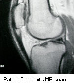 The investigation of choice for patellar tendonitis is an MRI scan. Specifically the MRI scan should include sagittal-stir sequences which demonstrate the high signal degenerative area in the deep surface of the tendon in the central area of the tendon in close apposition to the inferior pole of the patella. Failure to undertake this particular sequence may result in the condition being overlooked.
Ultrasound can also be useful in this condition and demonstrates a hypo-0eccogenic area in the thickened tendon. However the technique is very much operator dependent and not usually well interpreted by the operating surgeon.
Occasionally other conditions can be associated with patellar tendonitis including diabetes, gout, para-hypothyroidism. Other conditions which can be a cause of anterior knee pain are commonly confused with patellar tendonitis. These conditions include, synovial plicae, chondromalacia, quadriceps tendonitis, patello-femoral subluxation and hyper-pressure, fat pad impingement or Hoffa's syndrome and patello-femoral arthritis.
Initial Management / Steroids and Injections
The initial management of patellar tendonitis is relative rest with avoidance of those activities which cause the symptoms. This may mean a restriction in jumping activities or jogging particularly on hills. Non impact sporting activities such as cycling or swimming are usually less troublesome are not associated with symptoms. Anti-inflammatory medication is usually helpful to alleviate the symptoms. The condition is commonly associated with tight hamstring muscles and these must be stretched out in all cases. There may also be some restriction to spinal flexion and to calf flexibility. Any associated foot pronation (flat foot deformity) should be corrected with insoles or foot orthotics. Physiotherapy is of some benefit and commonly used in the initial treatment of this condition. It has been variously reported to be useful but there is little objective evidence for this. Physiotherapy modalities usually include stretching, massage, muscle specific strengthening ultrasound or other electrical therapy. It has been suggested that eccentric lunging type exercises may be a helpful form of physiotherapy. Mr. Johnson is however sceptical of the effectiveness of such lunging exercises in alleviating the symptoms.
Steroid and local anaesthetic injections to the tendon or it's environment is not advised. The tendon carries a huge force during weight bearing and steroid injections can be associated with causing degenerative changes in the tendon and repeated injections have been reported to be associated with tendon rupture. This is a very serious complication requiring reconstructive surgery that often results in a poor functional outcome. Therefore except in unusual and special circumstances, steroid injections for this condition is not recommended. Mr. Johnson has recently come across patients being injected with a "sclerosant" which includes phenol. This cannot be recommended and theoretically may be associated with a high risk of tendon necrosis or rupture.
If despite non-operative treatment, modification of activity, anti-inflammatory medication, stretching, physiotherapy and orthotics if appropriate, the condition continues to be troublesome and restrict activity then operative treatment may be considered.
Non-operative Management
 Non-operative management in patients with modest problems or in patients who wish to avoid surgery usually includes recording a detailed history of the condition such as the details of the current symptoms and disability, current and previous medications and past medical history. Then a clinical examination is undertaken by Mr. Johnson and subsequently radiographs or MRI scans can be arranged if necessary. An opinion is usually then available and discussed with the patient. Mr. Johnson will outline options for further treatment, and patients will then be encouraged to participate in the decisions about which treatment options are most appropriate. Initial management for some patients may include physiotherapy, use of anti-inflammatory medication (Ibruprofen, diclofenac, vioxx etc), an infra-patellar strap. Injections either with methyprednisiolone or sclerosant are not usually recommended. Alternately if the symptoms are significant or non-acute or non-operative treatment has not been sucessful, the prospect of early surgical intervention and arthroscopy may be appropriate and this will be discussed with patients.
Initial non-operative management may include physiotherapy, use of anti-inflammatory medication (Ibruprofen, diclofenac, vioxx etc), but If this proves unsatisfactory with an inability to return to sporting activity then the surgical option can always be revisited.
<BACK to Indications / Contra - indications | NEXT: Surgery>
Related Links..
+ How to make an appointment
+ Patella Tendonitis - see all links
+ Patient Information home
+ See the clinic
+ More about Mr Johnson
+ top
© The Bristol Orthopaedic & Sports Injury Clinic 2003. privacy | contact | Powered By Create Medical